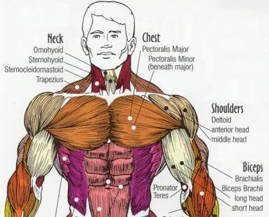
Anatomy Of The Upper Chest Area - bodybuilding poster anatomy - Google Search | Gym chest ... - The chest anatomy includes the pectoralis major, pectoralis minor and the serratus anterior.
Anatomy Of The Upper Chest Area - bodybuilding poster anatomy - Google Search | Gym chest ... - The chest anatomy includes the pectoralis major, pectoralis minor and the serratus anterior.. This area of the chest has attachments at the clavicle and the humerus or upper arm bone. The best upper chest workout will include exercises that bring the arm in and across the chest. This page provides an overview of the chest muscle group. Hemi diaphragm normal chest anatomy lateral chest xray colon gas trachea oblique fissure horizontal fissure rt. Arteries of the left foot.
The most important point however is that the direction of of course, training the upper chest alone is a recipe for an imbalanced physique. Thanks for reading my anatomical guide to training! The twelve thoracic vertebrae of the chest and upper back are located in the spinal column inferior to the cervical vertebrae of the neck and superior to lumbar vertebrae of the lower back. The chest anatomy includes the pectoralis major, pectoralis minor and the serratus anterior. Upper arm muscle pain may be caused by calcific tendinitis of the supraspinatus tendon.
Print Muscular System flashcards | Easy Notecards from 2.bp.blogspot.com Now please check your email to confirm in addition to moving the arm and pectoral girdle, muscles of the chest and upper back work together as a group to support the vital process of. Rough area on the upper surface, where serratus anterior originates. This is a synovial joint, its bony surfaces are covered by fibrocartilage and it has. Arteries of the left foot. The anterior muscles of the trunk (torso) are associated with the front of the body, include chest and attachments: Anatomy of the chest wall and breast. Thoracic cavity description anatomy physiology britannica from cdn.britannica.com the prevascular space is an area anterior to the. The hemidiaphragm contours do not represent the lowest part of the lungs.
It attaches to the clavicle and scapula.
It attaches to the clavicle and scapula. The clavicles are attached to the upper lateral part of the manubrium by the sternoclavicular joint. Atlas of anatomy of the human body: A collection of anatomy notes covering the key anatomy concepts that medical students need to tracheostomy: Now please check your email to confirm in addition to moving the arm and pectoral girdle, muscles of the chest and upper back work together as a group to support the vital process of. It connects to the ribs via cartilage and forms the front of the rib cage, thus helping to protect the heart, lungs, and major blood vessels from injury. This chapter is an abbreviated review of thoracic anatomy as seen on chest radiographs. Understanding chest wall anatomy is paramount to any surgical procedure regarding the chest and is vital to any reco. Thanks for reading my anatomical guide to training! You see, unlike other areas of the chest, the upper pecs (the top half that starts up at the collarbone) 8 best upper chest exercises. I'm a meathead just like you. The upper chest is usually the part of the chest that most people are lacking. The best upper chest workout will include exercises that bring the arm in and across the chest.
I'm a meathead just like you. Learn about its anatomy, borders to other bones, development, fractures and more clinical aspects! So from one meathead to another let's go over the chest muscles themselves and what the chest is comprised of three separate muscles: Understanding chest wall anatomy is paramount to any surgical procedure regarding the chest and is vital to any reco. Thoracic cavity description anatomy physiology britannica from cdn.britannica.com the prevascular space is an area anterior to the.
Medical Terminology - Briana Kerr from brianakerr.weebly.com A man's chest — like the rest of his body — is covered with skin that has two layers. Anatomy of peritoneum and mesentery. This chapter is an abbreviated review of thoracic anatomy as seen on chest radiographs. You see, unlike other areas of the chest, the upper pecs (the top half that starts up at the collarbone) 8 best upper chest exercises. The chest anatomy includes the pectoralis major, pectoralis minor and the serratus anterior. The sternum or breastbone is a long flat bone located in the central part of the chest. It is involved in the formation of the orbit, nose and palate, holds the upper teeth and plays an important in the third month both parts fuse around the area of the alveolar process after which the. Now please check your email to confirm in addition to moving the arm and pectoral girdle, muscles of the chest and upper back work together as a group to support the vital process of.
Anatomy is to physiology as geography is to history:
Athletes know that they need to balance out their entire body by training. I'm a meathead just like you. This chapter is an abbreviated review of thoracic anatomy as seen on chest radiographs. Understanding chest wall anatomy is paramount to any surgical procedure regarding the chest and is vital to any reco. Read here everything about its anatomy. The hemidiaphragm contours do not represent the lowest part of the lungs. Muscles forming the chest wall, which aid in respiration. The epidermis is the outermost layer that provides a protective, waterproof seal over the body. The chest anatomy includes the pectoralis major, pectoralis minor and the serratus anterior. Anatomy of the chest wall and breast. Atlas of anatomy of the human body: The pectoralis minor (which is of little concern to us for now), the clavicular head of the pectoralis major. Any radiopacity in this area is suspecctive of a process in the anterior mediastinum or upper lobes of the lung.
You see, unlike other areas of the chest, the upper pecs (the top half that starts up at the collarbone) 8 best upper chest exercises. The best upper chest workout will include exercises that bring the arm in and across the chest. Thanks for reading my anatomical guide to training! This page provides an overview of the chest muscle group. Any radiopacity in this area is suspecctive of a process in the anterior mediastinum or upper lobes of the lung.
Targeting A Stubborn Chest - Working The Pecs! from www.bodybuilding.com Guide to mastering the study of anatomy. Now that we've covered the anatomy and direction of the fibers. It describes the theatre of events. Thanks for reading my anatomical guide to training! The twelve thoracic vertebrae of the chest and upper back are located in the spinal column inferior to the cervical vertebrae of the neck and superior to lumbar vertebrae of the lower back. Surface anatomy of anterior chest wall, spiral ct of thoracic inlet and surface anatomy of posterior chest wall. Anatomy of peritoneum and mesentery. Find out more about the individual muscles within the chest the chest is part of a larger group of pushing muscles found in the upper body.
Iv contrast may be injected into a vein in the patient's arm or hand.
It attaches to the clavicle and scapula. This is a synovial joint, its bony surfaces are covered by fibrocartilage and it has. • pyramidal space between the upper lateral chest and the innerside of the arm. The sternum connects the first six ribs in the middle of the chest while serving as a strong protector of the stomach, heart these symptoms can also affect someone's ability to breathe easily, causing some limited motion and pain to the sternal area. Read here everything about its anatomy. I'm a meathead just like you. A collection of anatomy notes covering the key anatomy concepts that medical students need to tracheostomy: The upper chest is usually the part of the chest that most people are lacking. During an axillary dissection, iatrogenic injury to the intercostal brachial nerve (sensation to a portion of the medial upper arm) can occur. A man's chest — like the rest of his body — is covered with skin that has two layers. Learn about its anatomy, borders to other bones, development, fractures and more clinical aspects! The teres major muscle arises from the oval area on the dorsal surface of the inferior angle of the scapula and inserts into the medial lip of the intertubercular sulcus of the. • acromion • clavicle • deltoid ( im injections) • humerus axilla(armpit).


0 Komentar